


Use of synthetic hydroxiapatite and MTA in periapical surgery: A case report
Tatiana Teixeira de Miranda, Leonardo Rodrigues, Angélica Cavalheiro Bertagnolli, Alexsander Ribeiro Pedrosa, Carlos Henrique Martins de Oliveira
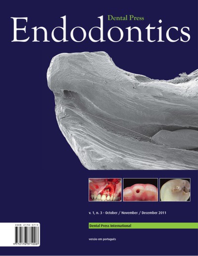
Objective: This article aimed to report a case of periradicular surgery in which biomaterials, such as MTA and synthetic hydroxiapatite were used. A periapical radiograph showed an extensive radiolucent area extending from the mesial aspect of the tooth 21 to distal aspect of tooth 22. Apicoectomy was performed and root-end cavities were prepared and restored with MTA as a retrofilling material. Synthetic hydroxiapatite was also used aiming to model the lost bone structure. The enucleated lesion was submitted for histopathological examination. A diagnostic of periapical granuloma was established based on the microscopic analysis. Two years after the periradicular surgery, there were no clinical or radiograph suggestive signs of treatment failbure. Instead, the patient’s follow-up has shown that the case management has been successful as indicated by lesion regression and periodontal repair. Based on this case, we can conclude that the definitive diagnosis of the type of periapical lesion can only be made by a histological examination and apical surgery can be an excellent complementary procedure when endodontic treatment has not yielded healing outcome.
Keywords: Periapical diseases. Periapical periodontitis. Periapical tissue.
How to cite: Miranda TT, Rodrigues L, Bertagnolli AC, Pedrosa AR, Oliveira CHM. Use of synthetic hydroxiapatite and MTA in periapical surgery: A case report. Dental Press Endod. 2011 Oct-Dec;1(3):51-5.
Wednesday, February 05, 2025 05:50